Published Nov 11, 2026



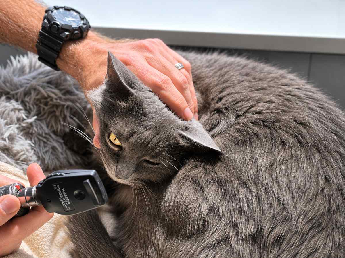
An ophthalmoscope is an important tool your vet uses to diagnose a variety of eye disorders in cats. Chris Vanderhoof / Cats.com
Keratoconjunctivitis sicca (KCS), also commonly termed “dry eye,” is an uncommon condition in cats but very important to recognize and diagnose, as it causes pain and can affect vision. In this article, you’ll learn some ways to recognize more serious eye problems in cats, how veterinarians diagnose dry eye, and how it’s treated.
Quick Overview: Dry Eye (Keratoconjunctivitis Sicca) in Cats
 Other Names: Dry eye, dry eye syndrome (DES)
 Common Symptoms: Thick mucus-like build-up on the eye surface, redness on the eye surface, constant scratching or rubbing the eye
 Requires Ongoing Medication: Yes
 Vaccine Available: FVRCP/HCP vaccine that includes prevention for feline herpesvirus (FHV-1) may reduce the risk of herpesvirus as an underlying cause for cats
 Treatment Options: Artificial tears and lubricants, topical ophthalmic cyclosporine, topical ophthalmic tacrolimus. Surgical relocation of the parotid salivary gland duct to provide lubrication to the eye with saliva is uncommonly needed but the only approach left if medical therapy is not successful.
 Home Treatment: Always keep rechecks with your veterinarian or veterinary ophthalmologist. These are extremely important to determining the right dose and frequency of eye medications to manage this condition and keep your cat comfortable. Eye medication administration may need to be frequent, especially for severe cases of dry eye. Ask your veterinarian or veterinary ophthalmologist for the best balance of medicating around your daily schedule. Administer eye medications with your cat facing away from you and tucked between your legs on the floor or a seat to keep them from backing away. A towel wrap may also be helpful as shown in our How to Give Cats Liquid Medicine Treating a dry eye condition requires life-long management. It’s important to be aware that reducing or stopping medications when things look better will often cause them to worsen again.
What Is Dry Eye (Keratoconjunctivitis Sicca)?
The term keratoconjunctivitis sicca originates from kerato- referring to the cornea or surface of the eye, conjunctiv- referring to the tissues beneath the eyelids and covering the whites of the eyes (sclera), and -itis referring to inflammation. Sicca is a medical term derived from Latin, meaning “dry.” Thus, keratoconjunctivitis sicca literally means an inflammation of the cornea and surrounding eye tissues due to dryness.
KCS, or dry eye as it’s commonly called, occurs when one or more components of the tear film that lubricates and protects the surface of the eye are deficient or do not function properly.
The surface of the eye, the cornea, does not have its own blood supply and so relies on the production of tear film for lubrication, protection from debris, and for sustaining nutrients like oxygen.
There are three components to the tear film, which are all produced by different areas around the eye surface. The most important layer is the aqueous (water soluble) layer, which is the thickest layer and most important for providing nutrition to the eye surface. This layer is produced by glands called lacrimal glands. These glands are located in the upper eyelid as well as the third eyelid (that extra protective lid that we humans don’t have but our furry friends do).
There is a lipid (fat) layer that is present on top of the aqueous layer. The lipid layer prevents the aqueous layer from evaporating, and is produced by the meibomian glands located all along the eyelids.
The third layer resides beneath the aqueous layer and is called the mucoid layer. This layer’s job is to create surface tension and allow the other aqueous layer to distribute evenly across the eye surface. The mucoid layer is produced by cells called goblet cells in the conjunctiva, which reside beneath the eyelids.
While KCS can be caused by any loss of the three layers, loss of production of the aqueous layer is the most common and the most harmful. Without it, the eye surface loses moisture and nutrition.
Causes of Dry Eye in Cats
There are a couple of underlying causes for dry eye in cats.
One of the most common causes occurs when the immune system attacks the lacrimal glands, preventing them from working. A genetic predisposition may exist for some cats.
Other causes include:
- Herpesvirus (FHV-1) infection that leads to keratitis (inflammation of the cornea) may result in dry eye as well from causing inflammation of the lacrimal glands.
- The sulfa class of antibiotics, which includes trimethoprim sulfa, has been connected with the development of dry eye. Fortunately, this is an uncommonly used antibiotic in cats.
- While uncommon in general with cats on these medications, non-steroidal anti-inflammatory (NSAID) medications have also been connected to development of dry eye.
- A condition where the nerves that stimulate the tear glands do not function properly, called neurogenic KCS.
- Hypothyroidism, while extremely rare in cats, has been connected to dry eye.
Symptoms of Dry Eye
Dry eye in cats can have some varying appearances depending on how serious it is.
At its worst, KCS will cause a cat to have a red, irritated looking eye with a thick mucousy discharge. This occurs because the eye has lost moisture and nutrition. The heavy mucousy discharge occurs because in most cases, the other components of tear film, the lipid and mucoid layers, are still being produced, but not the aqueous layer. This makes the tear film thick and kind of sticky.
The loss of protection and moisture to the surface of the cornea can lead to a condition called corneal ulceration. Loss of health and protection for the cornea can lead to bacterial infection, making a corneal ulcer worse. While KCS is already uncomfortable, a corneal ulcer makes things especially painful.
One exception to this type of ulcer is when feline herpesvirus is causing a herpetic ulcer on its own. This type of ulcer, also called a dendritic ulcer, may be present if FHV-1 is an underlying cause for dry eye.
Dry eye can share appearances with other eye conditions, making it very important for your veterinarian to provide the diagnosis. Corneal ulcerations, for example, may also occur secondary to a wound or scratch on the eye or abnormalities to a cat’s eyelashes or eyelids that cause corneal irritation and damage.
Complications of Having Dry Eye
If dry eye is not properly diagnosed, it does cause chronic, ongoing pain for your cat.
But looking even further down the road, dry eye has consequences for vision. Over time, chronic irritation to the cornea leads to cloudy scarring and pigmentation (a brown staining to the cornea) that can obscure vision. Pigmentation may be reversed to a degree with treatment and help with improving vision but often cannot be fully resolved.
Diagnosis of Dry Eye in Cats
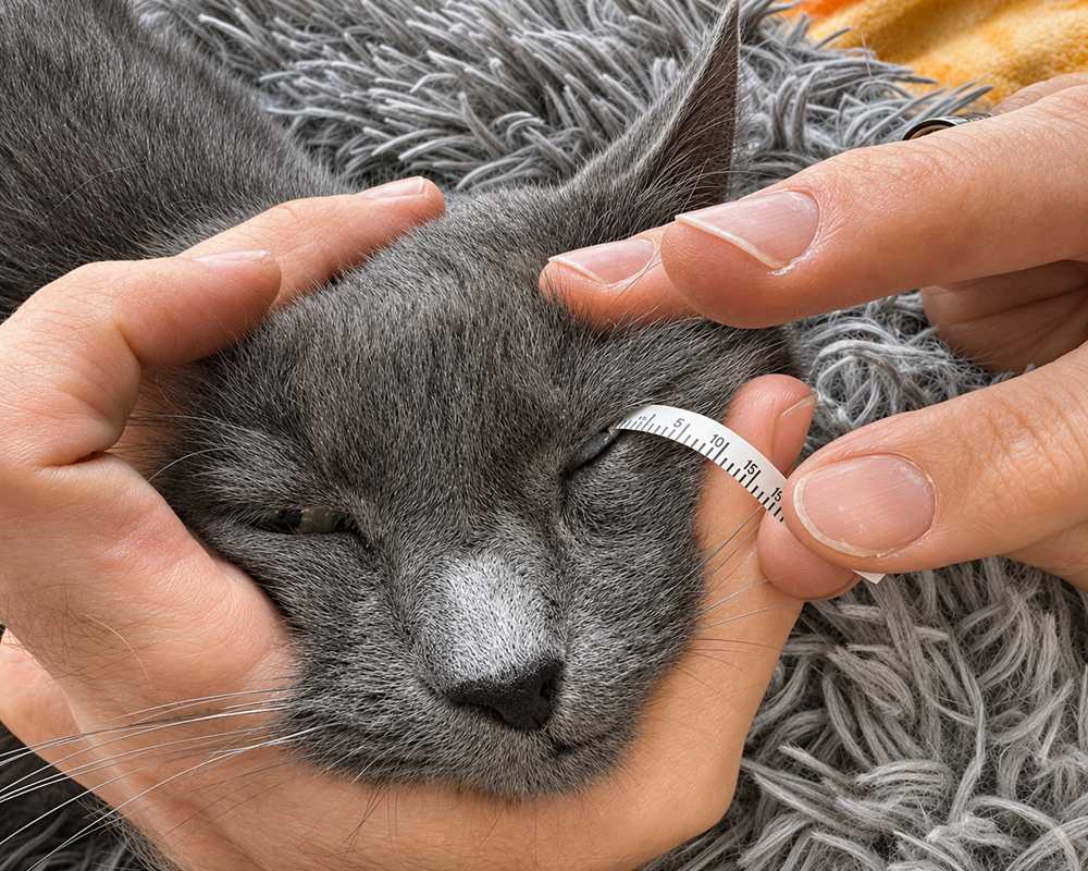
The Schirmer tear test (STT) uses a strip of absorbent paper placed beneath the lower lid. It assesses tear production by measuring in millimeters how much tear moisture the strip soaks up over the course of a minute. Chris Vanderhoof / Cats.com
The Schirmer tear test (STT) uses a strip of absorbent paper placed beneath the lower lid. It assesses tear production by measuring in millimeters how much tear moisture the strip soaks up over the course of a minute. Chris Vanderhoof/Cats.com
KCS is diagnosed using a test called a Schirmer tear test (STT). This test involves inserting a small strip of paper with a number scale printed on it just under the lower lid. The numbers are in millimeters. The strip of paper absorbs tears from the surface of the eye. The progression of the tears soaking into the paper strip is measured, usually over the course of one minute.
In dogs, a common normal range for a Schirmer tear test is 15-20 millimeters over one minute.
But for cats, this result can vary. When stressed, cats may not produce ample amounts of tears, artificially lowering their STT result. Thus, the results of the test must be interpreted in light of the cat having signs consistent with dry eye.
Another test called tear film break-up time may be helpful in supporting a potential diagnosis of dry eye. This test involves applying a drop of fluorescein dye (the same dye used to diagnose corneal ulcers) to the surface of the eye. The lids are closed a couple times to allow the dye to disperse across the surface of the eye. The dye is then closely observed under a special light source using a cobalt blue light filter (which looks similar to a blacklight).
The dye’s dispersion across the surface of the eye can be observed to separate over time where it loses its even distribution. In a normal eye, it should take 60 seconds or longer for this to happen. In a pet with dry eye, this occurs in a much shorter period of time.
Your veterinarian may choose to do a couple additional eye tests at this time. While not tests for dry eye itself, these other tests can help to rule out any other contributing causes or other eye abnormalities at the same time.
Fluorescein dye test (FDT): this test, involving application of fluorescein dye to the surface of the eye just like for tear film break-up time, is specifically for detecting the presence of a corneal ulcer. Corneal ulcers can occur at the same time as KCS and are very important to be aware of as treatment may be altered until an ulcer heals. This test may be done at the same time as the tear film break-up time, which also uses fluorescein dye. Unlike with tear film break-up time, using fluorescein dye to check for ulcers involves flushing the dye out using an eye wash, to see if any dye was taken up by an ulcer in the cornea and remains visible with the cobalt blue light.
Tear duct drainage: this test may also be done concurrently with fluorescein dye to see if tear drainage is occurring properly. If the tear ducts are functioning, dye should start to be visible just at the opening of the nostrils within 5-10 minutes.
Intraocular pressure: this test assesses pressure inside the eye using a device called a tonometer. High pressure may be consistent with glaucoma, which can also cause an abnormal appearance to the eye surface. Low pressure may be associated with inflammation inside the eye, or uveitis, which may also contribute to an abnormal eye appearance.
Treatments for Dry Eye in Cats
One of the most important immediate treatments for dry eye in cats is eye lubrication. Even when other medications are started, using an eye lubricant is extremely important. Not all lubricants and artificial tear solutions are created equal and there are a couple that ophthalmologists tend to favor. These include Genteal tears, Systane, and Optixcare. A variant of Optixcare containing hyaluronin is also advocated.
The frequency with which eye lubricants must be given depends greatly on the severity of dry eye and the condition’s response to other medications. Initially for severe dry eye, a topical lubricant may need to be given hourly. When started on medications we’ll review below, a common frequency for eye lubricants is every four hours. This frequency may be further reduced depending on the response to other medications to stimulate tear production.
There are two other primary medications for treating dry eye. In both cases, their goal is to stimulate aqueous tear production. As many cases of dry eye are immune-mediated, utilizing these immune-modulating medications helps to suppress that impact on the lacrimal glands and allow them to increase production of tears:
Cyclosporine (Optimmune): this is often the first medication started. It commercially only comes in an ointment form at a 0.2% strength and is typically started by applying a ¼-½ inch strip to the eye surface every 12 hours. A recheck of tear production 2-3 weeks after starting can be used to determine if the frequency of administration can stay the same or may need to be increased. The concentration may also sometimes require being increased (usually to 1-2%), but in that case, must be formulated through a compounding pharmacy. Non-ointment forms may also be formulated through a compounding pharmacy, usually in an oil suspension, in the event that the ointment form is harder to administer. About 80% of patients will respond to cyclosporine alone.
Tacrolimus is another immune-modulating medication that is used as a lacrimostimulant (tear production stimulator). There is no commercially available form of tacrolimus for the eye. All forms used for this purpose must be formulated with a compounding pharmacy. A 0.03% oil suspension is most common. Tacrolimus is most often added on top of using cyclosporine in cases where improvement with cyclosporine is not enough to achieve normal tear production. Some ophthalmologists do prefer to start with tacrolimus instead.
Side effects to these medications given topically are uncommon but the most common adverse effect is eye irritation, which occurs in about 5% of patients. In this case, the vehicle (meaning the oil type in a liquid suspension) may be changed or if the medication itself is suspected, may need to be discontinued in favor of adding in or adjusting the second.
Pilocarpine is a medication used to treat neurogenic KCS only, where the underlying cause is not the lacrimal glands themselves but instead the nerves that allow them to function properly. Pilocarpine may be given topically to the eye or the ophthalmic preparation may also be added on top of food and ingested orally.
In nearly all cases, medical therapy with eye medications must continue for the duration of a cat’s life in order to manage dry eye and keep it under control. Treatment may be more intensive at first, especially for more severe cases, but the hope in some cases is that medication frequency may be reduced if there is a good response.
There are a couple of other medications that may also be needed during dry eye treatment, though their use is temporary.
Ophthalmic antibiotics: these are needed if a corneal ulceration is present, which can be determined using the fluorescein dye test discussed earlier. Ofloxacin is a common choice among ophthalmologists, but others may be chosen. A corneal cytology or bacterial culture may also be elected to determine the correct antibiotic to use. An antibiotic must be continued until the ulcer fully heals.
Ophthalmic steroids: the use of topical steroids for KCS like prednisolone acetate is debated but may be utilized if deemed safe and appropriate. A topical steroid may help to reduce inflammation on the corneal surface and help initially stimulate the lacrimal glands to produce tears. However, steroids cannot be used if an ulcer is present in the eye, as this will cause the ulcer to worsen. Practitioners may also worry that even if they don’t find evidence of an ulcer initially that one may develop while the patient is receiving a topical steroid, making the condition rapidly develop and worsen.
Oral/systemic steroids would be considered ineffective for conditions affecting the cornea, as the cornea does not receive blood flow, depending on tears for its health and nutrition.
Surgery: While not performed commonly, a specific surgery may be performed in cases where medical management of dry eye with topical eye medications fails. The surgery is called a parotid duct transposition and involves relocating the duct for the parotid salivary gland that sits on either side of the face. Instead of opening into the mouth, the duct is redirected to open at the corner of the inside of the lower eyelid.
While saliva is the body’s best alternative to tears and the success rate for the procedure is about 90%, calcium mineral deposits often occur over time and a pet will require additional eye medications to manage the side effects. This procedure is typically only performed by a veterinary ophthalmologist specially trained in the procedure and will help tailor additional therapies depending on the patient’s needs.
Surgical removal of an eye, called enucleation, is not a result we hope is ever needed. However, a reality is that progression of dry eye if not treated properly will eventually lead to an eye that is permanently blind and chronically painful for the kitty. Vision by this time cannot be restored and surgical removal of the eye helps to relieve that discomfort for the long term. This procedure is considered an end-stage or salvage procedure designed only to relieve chronic discomfort from a painful, non-functional eye, and can be performed by many general practice veterinarians.
Cat Care Tips
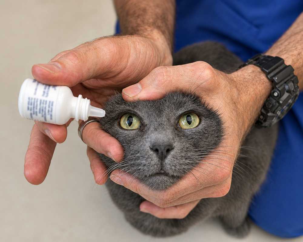
Position your cat facing away from you to help prevent backing up while you administer eye medication. Chris Vanderhoof / Cats.com
- When giving eye medications, it helps to have your cat face away from you, either in a seat or on the floor, positioned between your legs. This way, as you administer the eye medication, they cannot back up on you. Some cats may benefit from being wrapped in a towel or blanket, similar to what may be needed for giving an oral medication.
- The frequency of eye lubricants needed may be difficult to do with the average person’s schedule. While giving them every four hours spread evenly throughout the day may not be possible unless you happen to work from home, you can adopt a schedule of applying them first thing in the morning, right before leaving the home, right when you get home, and then late before bed.
- Always make sure to keep recheck appointments scheduled with your veterinarian or veterinary ophthalmologist. Especially early on, it will not be clear how effective an initial medication dose and frequency will be for your cat. These rechecks are crucial to see if any adjustments to the medication concentration or dosing frequency are needed. These rechecks are also needed to see if adding in a second medication, such as adding tacrolimus on top of using cyclosporine, will be needed.
- Whenever using an ointment, like Optimmune, along with another non-ointment product like an artificial tears solution or oil suspension, always use the non-ointment product in the eye first. If the ointment is applied first, the second non-ointment product will not spread over the eye properly.
- When administering more than one eye medication at a time, it is best to separate the administrations out by 5 minutes. This allows time for one medication to spread over the eye before the second is added. However, if you have only one chance at a time to collect your kitty and administer eye medications (before they run and hide and are nowhere to be found), err on the side of getting your medications in instead of risking missing a dose.
Prevention of Dry Eye in Cats
Dry eye may not be fully preventable given its underlying causes often having a genetic component. Starting eye medications for dry eye before dry eye is actually present and diagnosed will not prevent the condition from developing if it was always going to.
Vaccinating properly for feline herpesvirus with the FVRCP/HCP vaccine can be one way to prevent and/or reduce the impact of feline herpesvirus causing herpetic ulcers and potential dry eye. Unfortunately, while vaccination is still recommended, some very young kittens may acquire FHV-1 prior to an appropriate age to start vaccination.
One potential cause of dry eye in cats, while uncommon, is entirely preventable. A prolapsed third eyelid or “cherry eye” as it is more often called, may occur in some young cats, usually due to a genetic predisposition. If you recall from early in the article, the third eyelid (or nictitating membrane as it is also called) contains lacrimal glands and is partly responsible for production of aqueous tears. An older and outdated method of treating cherry eye in dogs and cats was to simply excise or cut off and remove, the third eyelid.
While an apparent quick and easy fix to an unsightly problem, many studies and clinical experience has demonstrated that the loss of the lacrimal glands of the third eyelid carries a very high risk for developing dry eye within a couple of years. Unfortunately, these cases are harder to treat because the tear stimulating medications cannot work on the gland if the gland is actually absent. These patients depend highly on regular use of artificial tears and lubricants, or the parotid duct transposition surgical procedure for the best outcomes.
However, the good news is that this cause is entirely preventable, as third eyelid excision has fallen greatly out of favor as a treatment for cherry eye for this reason. Surgical approaches to replace the third eyelid gland back into its proper place through tucking, tacking, or imbrication, are far more preferable and can be performed by many veterinary general practitioners.
Medication-associated dry eye, such as with NSAID or sulfa medications cannot be predicted but are at least quite rare. The effects causing dry eye are often temporary and tear production usually returns once the medications are discontinued. This is not guaranteed to occur however and in cases where the effect is permanent, the treatment approaches described earlier may be used.
-
Barnes, C., Hunter, T., Downing, R., & Ward, E. (n.d.). Keratoconjunctivitis Sicca (KCS) or Dry Eye in Cats. VCA Animal Hospitals.
-
Brooks, W. (2024, November 6). Dry Eye (Keratoconjunctivitis Sicca) in Dogs and Cats. Veterinary Partner.
-
Geller, A. (2009, April 1). Feline Keratitis and Conjunctivitis. Dvm360.
-
Sebbag, L., Pesavento, P. A., Carrasco, S. E., Reilly, C. M., & Maggs, D. J. (n.d.). Feline dry eye syndrome of presumed neurogenic origin: a case report. Journal of Feline Medicine and Surgery Open Reports. https://doi.org/10.1177/2055116917746786
-
Fentiman, K. E., & McCool, E. S. (2025). Conjunctivitis and Keratitis. The Feline Patient, 177–181. https://doi.org/10.1002/9781394218035.ch55
-
Valley Central Veterinary Referral and Emergency Center, & Landis, M. (2015). Keratoconjunctivitis Sicca (KCS) and other Tear Film Abnormalities. Central Connection VCVREC Spring 2015 Newsletter.
-
Gionfriddo, J. (2010, October 1). Just Ask the Expert: What is a Normal Schirmer Tear Test Result. Dvm360.
-
eye vet. (n.d.). Parotid Duct Transposition. Eye Vet.
-
Brooks, W. (2025, January 20). Cherry Eye in Dogs and Cats. Veterinary Partner.